F379 (2017)
F379 (2017). Ultrafast elemental and oxidation-state mapping of hematite by 4D electron microscopy.
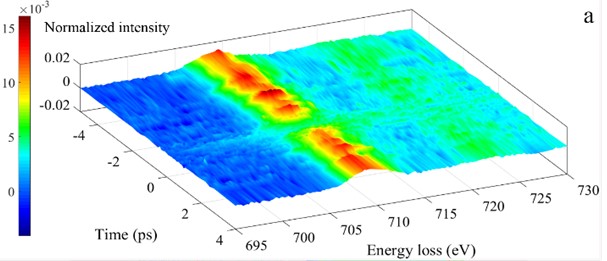
A new methodology sheds light on the fundamental electronic processes that occur at the subsurface regions of inorganic solid photocatalysts. Three distinct kinds of microscopic imaging are used that yield spatial, temporal, and energy-resolved information. The value of this methodology is illustrated by studying afresh a popular and viable photocatalyst, hematite, α-Fe2O3. By employing high-energy electron-loss signals (of several hundred eV), coupled to femtosecond temporal resolution as well as ultrafast energy-filtered transmission electron microscopy in 4D, we have, inter alia, identified Fe4+ ions that have a lifetime of a few picoseconds, as well as associated photoinduced electronic transitions and charge transfer processes. The experiments were performed by Dr. Z. X. Su at California Institute of Technology under supervision of Prof. Ahmed Zewail. [Z. X. Su, J. S. Baskin, W. Z. Zhou, J. M. Thomas, A. H. Zewail, J. Am. Chem. Soc. 139, 4916–4922 (2017).]