F221 (2005)
F221 (2005). Direct observation of defects in zeolite beta.
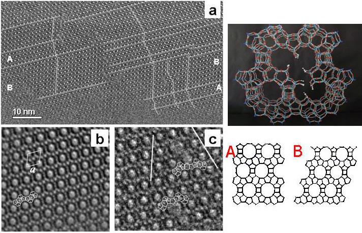
Left: (a) HRTEM image of an edge of a zeolite beta crystallite, annotated to show the stacking directions in different parts. Nanodomains related to polytpyes A and B (bottom right) are indicated.Defects are visible in the center of the image, where domains with different stacking directions meet. (b) Fourier-averaged image of a domain of type B, with structural units outlined. (c) Image of two double pore defects, showing different stacking directions and outlining secondary structural details on either side of the defects. Top right: Model of the observed defects, obtained by stacking in the two ways onto a single layer and continuing to the third similar layer. This work explains the location of the silanol groups commonly existing in zeolite beta. [P. A. Wright, W. Z. Zhou, J. Perez-Pariente, M. Arranz, J. Am. Chem. Soc. 127, 494–495 (2005)].